When I was eight years of age I was sent to hospital with what doctors described as “a troublesome foot”. The first diagnosis made of me was “Post Polio”. Just before I reached the age of ten, I underwent a series of brain operations which many people believe were “experimental” for a condition which medical records call “Dystonia Musculorem Deformens.”
Today my disability is called Generalised Dystonia or Primary Dystonia and is described as a “chronic involuntary movement disorder” for which there is no known cause and no known cure. Finding medical records relating to my time in hospital or records pertaining to surgery performed on me has proven very difficult. Eventually a few pages did surface – one reads as follows:
“Admitted for Stereotactic treatment of lower limb disc and dyskenesia.
An electrode was inserted into the Globus Pallidum but for technical reasons it was not possible to coagulate it. The Patient appeared to improve following insertion of the electrode” Another medical record reads: Readmitted for possible basal gangloin surgery. A small abscess had formed around one of the markers and therefore no surgery could done, but the marker was removed. Following an inquiry made regarding brain surgery carried out on me the following letter was received by a doctor who requested it on my behalf.
The record shows that this was a patient of Dr (Named) with a diagnosis of Dystonia Musculorum Deformans which was treated by an incomplete right stereotactic pallidotomy on the 6th February 1961. The planned electrocoagulation could not be completed because of a breakdown in the electrical apparatus. However, from the mechanical leesion alone, the patient seemed to have improved. I should add that the “several” operations you mentioned in your letter gives an inaccurate impression. There was only one operation but this was preceded and followed by minor procedures under local anaesthesia for the placing and removing of metallic markers used in the Stereotactic procedure.
This letter is signed by a neurosurgeon and refers to surgery carried out on me in 1960/61. I was 9 to 10 years of age.
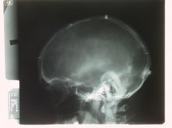
In February 2004 an orthopaedic surgeon suggested that I have an MRI scan done with a view to possible spinal surgery to alleviate chronic back and leg pain. Two days before the scan was due to be done under general anaesthetic, I happened to mention to staff in the radiology department that I might have metal in my body – possibly in my head. A decision was made to do an x-ray as a matter of urgency. There was an eerie silence in the x-ray department as a radiographer suggested that I should wait to see a radiologist. The radiologist informed me that and MRI scan would not be done because “there is what appears to be metal in your head”. I asked to see the x-rays. I realised the “picture” was the head of a 52 year old male but in a flash I was catapulted back to waking in a hospital bed following brain surgery – I was 9 years old. I was traumatised. My suspicion of metal in my head was now a reality. Half a century on, I was seeing what I had so often suspected but could never prove – until now.
I’ve been unable to find what these “bits and pieces” are. I’ve been unable to find out what they were used for or why they were left there. What I have been told by one expert in the field of neurosurgery was that it would “be dangerous to remove them at this stage”. Why I would put pictures such as these on a website? Answer: In the hope that someone will be able to tell me what the point of inserting these bits was and why were they left there? If you know the answer to either of these questions then perhaps you would contact me below.
Thank you.

